UCL Injury Treatment in Baton Rouge
What is the UCL?
The ulnar collateral ligament (UCL) is a ligament that runs through the inner side of the elbow, connecting one of the forearm bones (ulna) to the upper arm bone (humerus). The UCL supports the elbow in its full range of motion, allowing it to twist and bend with comfort and ease. It also stabilizes the elbow joint during the strenuous motion of an overhead throw.
How do UCL Injuries Occur?
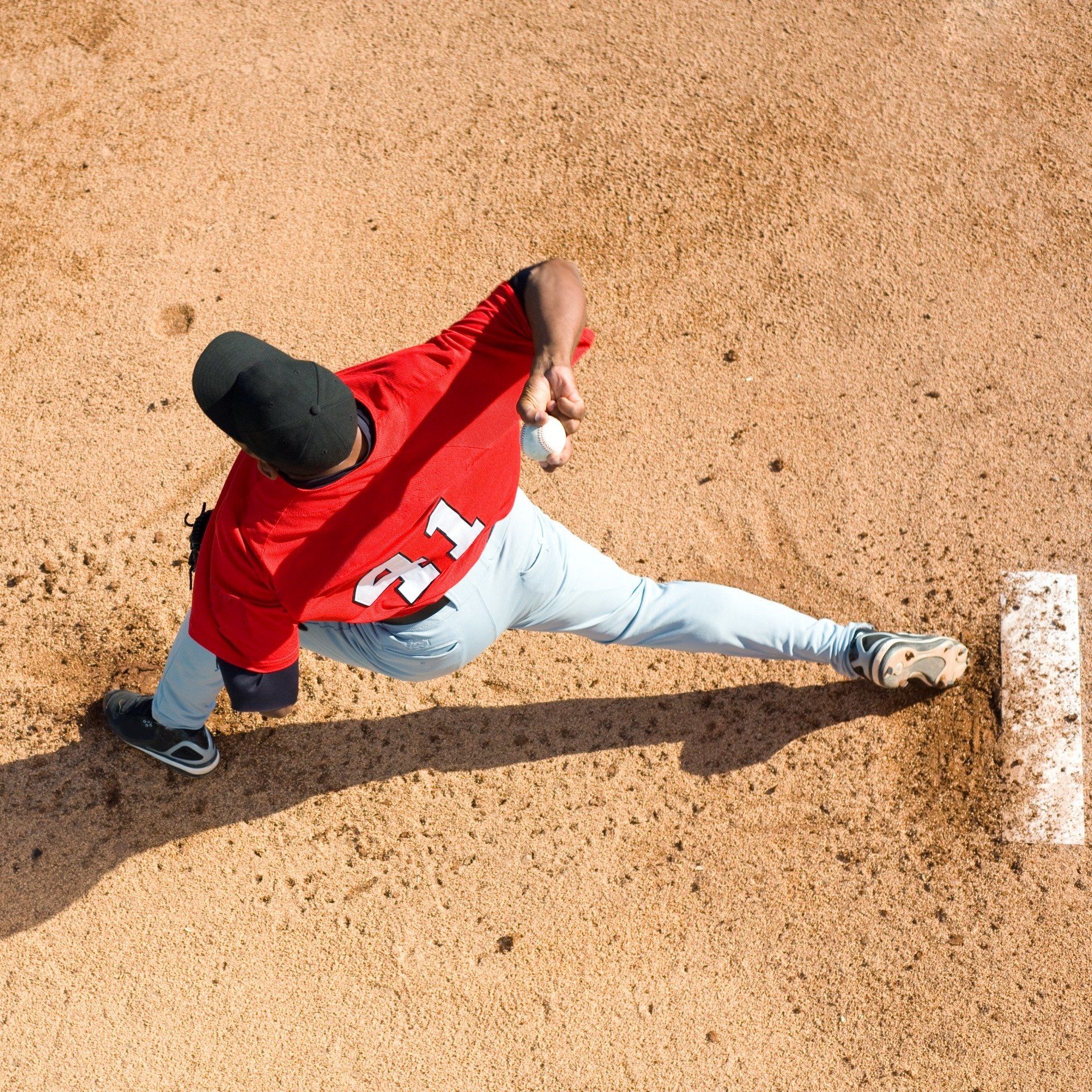
The overhead throwing motion, particularly when performed repeatedly, is the most common cause behind UCL injuries. This is why UCL-related problems are most common in athletes like baseball pitchers. However, they may occur in any individual who frequently uses the arm in a bending motion or through sudden incidents of trauma.
What are the Symptoms of UCL Injuries?
UCL injuries are typically chronic and occur gradually, over time. Less commonly, the onset of a UCL injury may be sudden, or acute, as can be the case with a fall or during athletic activities like gymnastics or wrestling. Symptoms of a possible UCL problem include:
- A sudden “pop” along the inside of the elbow
- Pain along the inside of the elbow
- Tenderness, bruising, or swelling at the elbow
- Elbow joint instability
- Weakened grip and throwing strength
How are UCL Injuries Diagnosed?
Diagnosing a UCL injury begins with a physical examination where the doctor will palpate the area to check for signs of ligament injury and test range of motion. One commonly used type of physical examination is the Valgus stress test where pressure is applied to the outside of the elbow to determine if the UCL can prevent the joint from opening up. If a sprain or tear of the ligament is suspected, an imaging test may then be ordered to confirm the diagnosis and identify the severity of damage. Tests commonly used, include:
- X-ray – Radiation is used to produce an image of the surrounding bones in an x-ray, helping rule out any potential fractures that may have occurred with the sprain.
- Magnetic Resonance Imaging (MRI) – MRIs produce more detailed images of soft tissue such as ligaments through the use of a magnetic field and radio waves.
- Ultrasound – Sound waves are used to help the physician judge the condition of the injured ligament directly as it is in different positions.
- CT Scan – In a CT scan, multiple x-ray images are taken from several different angles and used to create a 3D image with greater levels of detail
Grades of UCL Injuries
The severity of a UCL injury is classified according to grades. There are three different grades of injury:
- Grade I – This is the least severe level of sprain. Stretching of the ligament may cause pain, but there is no observed tearing.
- Grade II – This grade of sprain includes partial tearing of the ligament and is likely to produce significant amounts of pain and swelling.
- Grade III – This is the most severe level of sprain with complete tearing of the UCL. Grade III sprains will be accompanied by significant pain, swelling, and immobility.
Non-Surgical Treatment of UCL Injuries
Grade I and II UCL injuries are often successfully treated using non-surgical techniques. Some of these same techniques may also be used to ease Grade III injuries before surgery can be performed.
- R.I.C.E. –
Rest, ice, compression and elevation (R.I.C.E.) is the first line of treatment for injuries such ligament sprains for the first two to three days.
- Medication – Over the counter pain relievers like ibuprofen or acetaminophen can help control any pain associated with the injury.
- Mobility Devices – In higher grade sprains, a sling may be used to help immobilize the elbow, preventing additional problems and allowing the injury to begin to heal.
- Physical Therapy – As part of the healing process for more severe sprains, orthopedic physical therapy can help strengthen the damaged ligament, aid in elbow stability, and restore lost range-of-motion. A physical therapist can also help the patient learn better throwing mechanics to prevent future injury.
Tommy John Surgery for UCL Injuries
Tommy John surgery is the colloquial name for UCL reconstruction. This type of surgery is performed in the highest grades of UCL injury. During the procedure, the surgeon reconstructs the torn ligament by using a tendon either from the patient’s own body or from a cadaver. The most commonly used tendon in this surgery is the palmaris longus from the forearm.
Following Tommy John surgery, patients typically recover in mobility-limiting splint or brace for the first few weeks. This will be followed by a rehabilitative period of physical therapy that may last several months.
UCL Injury Specialists in Baton Rouge
ARTHUR E. HESS, M.D.
Pediatric & Adult Orthopedic Trauma
Hip, Knee & Shoulder Reconstruction
Hip Preservation
Total Joint Replacement Surgery
Robot-assisted total knee arthroplasty
General Orthopedics

MATHEW J. MAZOCH, M.D.
General Orthopedics
Sports Medicine
Shoulder Surgery

ALAN C. SCHROEDER, M.D.
Shoulder Arthroscopy
General Orthopedics
Orthopedic Sports Medicine
RELATED READING
UCL Injury Blogs